
Faced with the aging population and the rising prevalence of various chronic diseases, reasonable and scientific health risk assessment and management are helpful to better control personal and overall medical expenses and protect personal health interests to the greatest extent.
In recent years, the advantages of retinal image AI in health risk assessment have been shown. What advantages does it have? What value can it bring to the physical examination center and related departments?
At the Frontier Innovation and Wisdom Forum of the 9th 301 Discussion on Health and the 11th Summit Forum of Directors of National Physical Examination Center,Professor Tang Shiqi, deputy director of Health Management Branch of Chinese Medical Association, president of Hubei Health Management Society and academic leader of Health Management Center of Wuhan University People’s Hospital.Make a wonderful share with the title "Retinal Image AI Helps Health Risk Assessment".
In her speech, she pointed out:"Apart from the loss of life, there is nothing more frightening than the loss of vision. It is suggested that fundus photography should be added to chronic disease screening, eye health should be included in chronic disease system, fundus examination should be paid attention to, and the threshold should be moved forward to realize early detection and diagnosis of chronic diseases. "
The transcript of the speech is edited as follows:
Thank you very much for your introduction. The topic I share with you below is "Retinal Image AI Helps Health Risk Assessment". I will share it with you from four aspects, namely, the current situation of fundus diseases, retina as the best health assessment window, assessment of various diseases through retina, and the application of artificial intelligence products of retinal images.
Present situation of fundus diseases
"The 14th Five-Year National Eye Health Plan" listed fundus diseases as the key eye diseases in the next five years for the first time, and proposed early detection, early diagnosis and early treatment to reduce the blindness rate of diseases.
The burden of fundus diseases is heavy. There are more than 40 million patients with fundus diseases in China, and the distribution of medical resources is still uneven. At present, as the most important blinding eye disease in China, the clinical status of fundus diseases can be said to have a long way to go. This is also the main reason why our country has made fundus diseases a new disease in the National Eye Health Plan of the 14th Five-Year Plan released this year.
our countrydiabetesThe prevalence rate has reached 10%~13%, among which the prevalence rate of diabetic retinopathy in diabetic population is as high as 15%~43%. Then the risk of blindness in diabetic patients is 50~80 times greater than that in non-diabetic patients.
hypertensionIt is a common cardiovascular disease. The incidence rate in China is 5.11%, and about 70% of patients have fundus diseases. With the development of the course of the disease, changes such as hemorrhage, hard exudation, lint spots and the like appear in the fundus, and papilledema may occur in severe cases.
Age-related macular degeneration.For those of us over 50 years old, about one in every six people has macular degeneration. Early detection, diagnosis and treatment of macular degeneration are of great significance to protect central vision and prevent blindness. Therefore, it is recommended that people over 50 years old have a routine eye examination every year.
Fundus lesion caused by high myopia.There are about 70 million people with myopia greater than 600 degrees in China. It is recommended to screen the fundus every six months to one year.
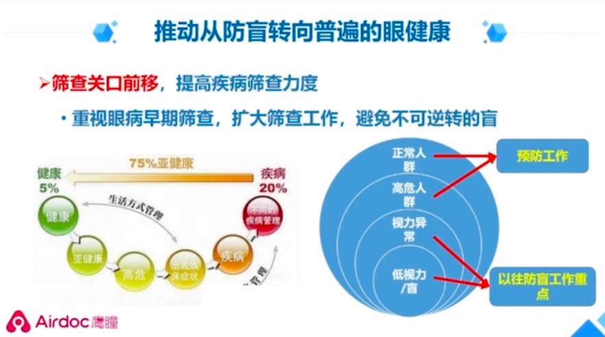
Therefore, we should promote the shift from blindness prevention to universal eye health, move the screening threshold forward, and improve the screening of diseases. At the same time, we should pay attention to the early screening of fundus diseases, expand the screening work and avoid irreversible blindness.
It is the best window for retinal health assessment.
There are very important structures in the fundus, including the optic nerve, retina, retinal arteries and veins, macular area, etc., and this is the area examined by the fundus.
Many fundus diseases have no obvious symptoms in the early stage and are difficult to be detected. However, the early stage is often the right time for treatment. Once missed, the lost vision may not be well recovered. 80%~90% of fundus diseases can be screened by taking a photo of fundus.
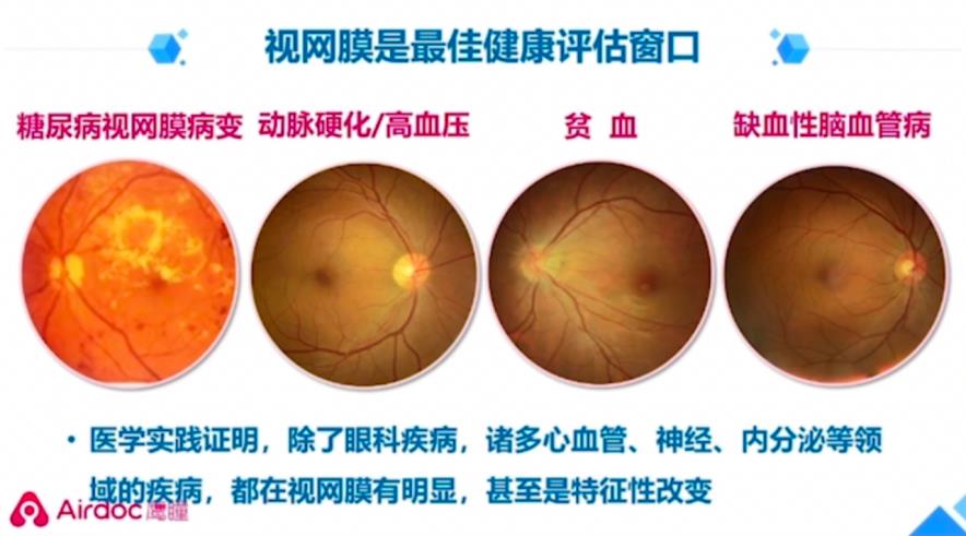
In addition, medical practice has proved that in addition to ophthalmic diseases, manyCardiovascular, neurological, endocrineOther diseases have obvious or even characteristic changes in the retina.
Evaluation of various diseases through retina
First of all.Diagnosis of diabetic retinopathy by retinal image AI. An SCI article published by Shibei Hospital, Jing ‘an District, Shanghai shows that artificial intelligence has high sensitivity and specificity in detecting sugar webs and proliferative sugar webs (the sensitivity and specificity of detecting sugar webs are 90.79%, 98.5%, AUC=0.946;; The sensitivity and specificity of detecting proliferative sugar net are 91.18%, 98.79%, AUC=0.950). It is feasible to carry out screening of sugar net based on artificial intelligence in community hospital outpatient department.
AI auxiliary diagnosis system for retinal diseases.This article of Zhongshan Eye Center of Sun Yat-sen University was published in the sub-issue of The Lancet, and its SCI impact factor was very high, reaching 24.519. Studies have confirmed that this "comprehensive intelligent diagnosis expert of fundus diseases" developed by Eagle Pupil Airdoc can identify 14 common fundus abnormalities, with an average AUC of 0.955, and it performs well in the screening of various fundus abnormalities.
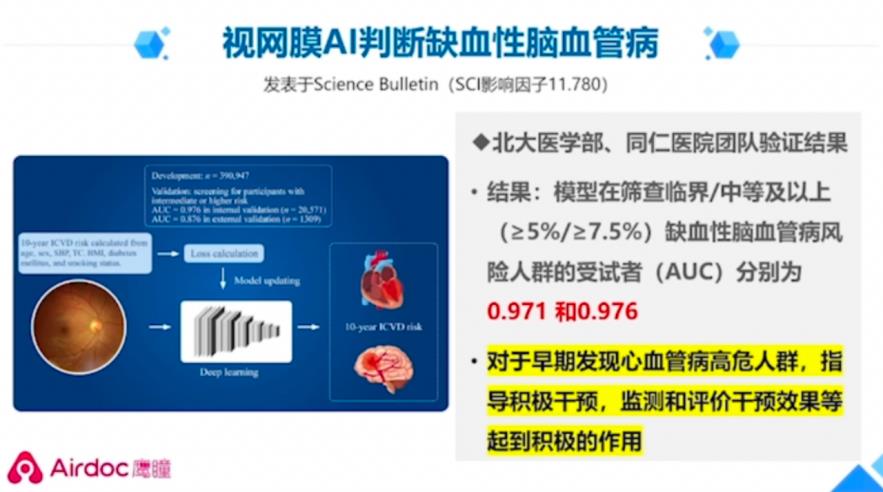
Retinal image AI to judge ischemic cerebrovascular disease.This is also a paper published in Science Bulletin, a journal sponsored by China Academy of Sciences, with an impact factor of 11.78. The results verified by the team of Peking University Medical School and Tongren Hospital show that (the model) plays a positive role in early detection of high-risk groups of cardiovascular diseases, guiding active intervention, monitoring and evaluating the intervention effect.
The traditional evaluation method of ischemic cerebrovascular disease has many collection indexes, so it needs invasive collection. The blood test takes a certain time, 2~4 hours, so the calculation is complicated and it is difficult to popularize. The new evaluation method based on China’s cardiovascular risk prediction model is retinal image +AI deep learning algorithm, which only needs fundus photography, non-invasive collection, and the detection time is 1 minute, and the report can be made directly in about 1 minute.
Retinal image AI can also evaluate Alzheimer’s disease.The retina is a part of our brain tissue. The degeneration of cranial nerves is also reflected in the subtle changes of retina. Taking CAIDE≥10 copies as the classification standard for high-risk groups (CAIDE dementia risk score is a risk assessment tool for the general population), the retinal AI algorithm is superior to the traditional invasive and complex dementia assessment, that is, it can be assessed by taking retinal photographs.
Retinal AI can also evaluate anemia.Retinal photographs of patients with anemia are usually pale or pale in background, pale in optic disc, and mild edema. The veins are pale. According to the model of retinal artificial intelligence, the AUC of anemia is > 0.9.
Application of retinal artificial intelligence products
Eagle Eye’s products are very good. It has multi-dimensional products and services. First of all, their artificial intelligence auxiliary diagnosis software can screen fundus diseases and lesions, make auxiliary diagnosis and assess the health risk of systemic chronic diseases, and has comprehensive qualifications, including the first three types of ophthalmology certificates in China, including the second, third and CE certificates.
Has excellent performance. The accuracy, specificity and sensitivity are leading in the world, and it is suitable for all mainstream fundus cameras in the market, including Canon and Topcom.
In addition, there is an authoritative endorsement. Carry out the world’s largest real-world research, with the results published in The Lancet magazine; Won the Wu Wenjun Prize twice, the highest award of artificial intelligence in China.
Eagle Airdoc’s fundus camera has the advantages of pupil-free, full-automatic, self-help (no operator required) and no professional darkroom. It is a self-developed domestic camera with high cost performance.
But also provide operational support services. Performance support, training support, follow-up of positive cases, and review by superior medical institutions, the Internet and hospitals.

(For the physical examination center), the retina image AI of Eagle Pupil Airdoc has seven values:
First,Upgrade the ability of eye examination.It can be used for auxiliary diagnosis of fundus diseases and risk assessment of systemic chronic diseases.
Second,Improve the standardization of diagnosis and treatment.It is a registered product, with standardized report, stable state, authenticity and reliability.
Third,Increase the efficiency of effective physical examination centers.
Fourth,Improve academic ability.It can quantify and multi-dimensionally empower scientific research, and artificial intelligence can improve the probability of project application, and expand clinical research opportunities of multi-departments and multi-diseases.
Fifth,Make up for the shortage of ophthalmologists.Automatic shooting, without doctor’s operation, AI-assisted diagnosis of liberation ophthalmologist. Nurses can easily operate it, and it is suitable for in-hospital examination and external examination.
Sixth,It is convenient to check in many occasions.The equipment is small, easy to operate, intelligent and fast, and it can be used in the out-of-home medical examination vehicle, which is suitable for large-scale screening.
Seventh,Historical comparison and continuous management.Realize automatic historical comparison of multiple inspections.
To sum up, apart from the loss of life, there is nothing more frightening than the loss of eyesight. It is suggested that fundus photography should be added to chronic disease screening, eye health should be included in chronic disease system, fundus examination should be paid attention, and the threshold should be moved forward to realize early detection and diagnosis of chronic diseases. A photo of the fundus, early knowledge of the disease.